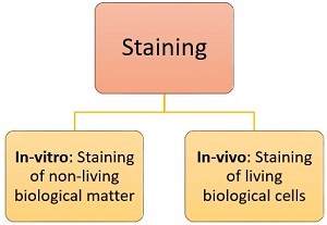
Describe How Staining Is Used on a Regular Basis
Mix well the culture with dye taken on the Glass slide. A large variety of fluorescent molecules are used on a regular basis to tag major histocompatibility complex MHC multimers for detection of antigen-specific T cells.
![]()
2 4 Staining Microscopic Specimens Microbiology Canadian Edition
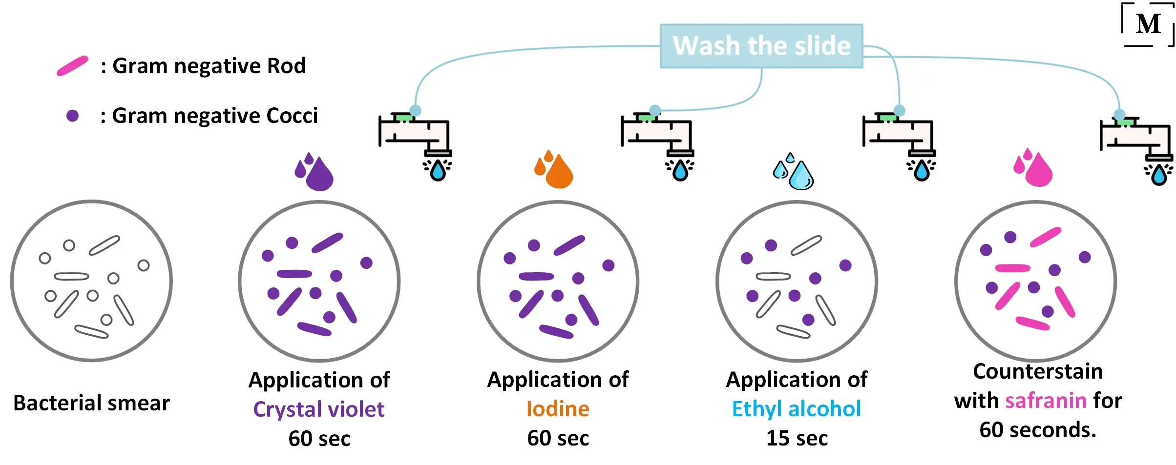
Finally a secondary stain safranin is added which counterstains the decolorized cells pink.

. The stain has been unchanged for many years because it works well with a variety of fixatives and displays a broad range of. It is referred to as a primary stain since it is applied first. This complex is a larger molecule than the original crystal violet stain and iodine and is insoluble in water.
A heat fixed smear is covered with a basic violet dye Example. Then after all cell appears red. A smear is subjected to heat after staining with Zeihl Neelson Carbol fuschin.
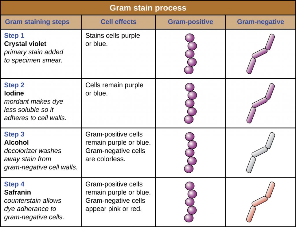
First cells are stained with crystal violet followed by the addition of a setting agent for the stain iodine. Then alcohol is applied which selectively removes the stain from only the Gram negative cells. The primary stain crystal violet binds to peptidoglycan coloring cells purple.
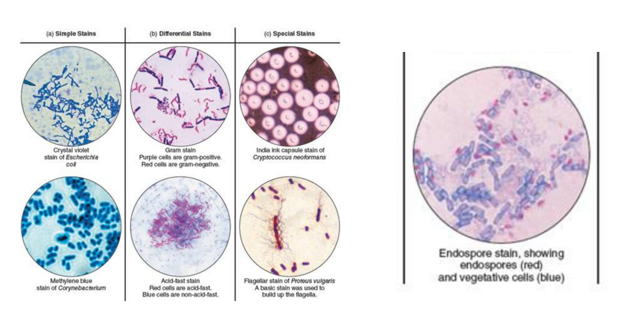
Positively charged cationic dyes such as methylene blue crystal violet safranin etc bind with negatively charged cellular constitutents such as nucleic acids and acidic polysaccharides and cell surface of bacteria. Hence we use dyes to stain cells. 1 Difference between simple and differential staining methods.
Fast stain differentiates an important group of bacteria the mycobacteria on the basis of lipid content of their cell wall. During steam carbol fuschin penetrates the bacterial cell. Based on the types and number.
Simple stain - A staining procedure involving only one stain that can be used to determine cell shape size and arrangement differential stain - A staining procedure involving two stains that can be used to differentiate one component or cellular structure from another or to differentiate an entity from another in a specimen. Now take another Microscopic Glass slide place it near to the specimen-dye mixture at an angle of about 30 45. When the smear is stained with carbol fuchsin it solubilizes the lipoidal material present in the Mycobacterial cell wall but by the application of heat carbol fuchsin further penetrates through lipoidal wall and enters into cytoplasm.
Grams Staining comprises of four steps. Both gram-positive and gram-negative cells have. The Gram stain involves staining bacteria fixing the color with a mordant decolorizing the cells and applying a counterstain.
INTRODUCTIONHematoxylin and eosin HE stains have been used for at least a century and are still essential for recognizing various tissue types and the morphologic changes that form the basis of contemporary cancer diagnosis. The process involves three steps. The different response of the two groups to the Gram stain is based on fundamental differences in cell wall structure and composition.
The cells of Mycobacteria species appear red whereas non-acid fast bacteria appear blue. Gram stain is used to differentiate the bacterial cells by staining the cell wall and distinguish two major groups of bacteria that are gram-positive and gram-negative. Capsular staining is a combination of two staining techniques.
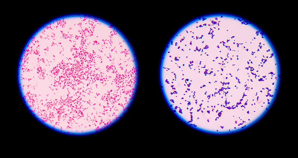
Gram-positive bacteria appear violet in colour and gram-negative bacteria appear pink in colour as a. 2 Theoretical and chemical basis of Gram stain procedure. We have evaluated the way in which the choice of fluorescent label can impact the detection of MHC multimer binding T cells in an ex.
It aids in the diagnosis of a specific organism and tells the difference between gram negative and gram positive bacteria. Acid-fast staining is based upon the principle of staining the bacterial cell relative to their cell wall differences. Are the basic dyes.
The smear is next decolourized by washing with ethanol or acetone. Gram positive and Gram negative. Principle of Acid-Fast Stain.
Methylene blue crystal violet safranin etc. Manevals stain to color the cell proper of the bacteria. 3 Relationship between Gram staining and physiology of the microbe 4 Role of chemicals used during the Gram staining procedure 5 Appearance of Gram positive cells and.
Congo red to color background Simple stain. Gram Staining is the common important and most used differential staining techniques in microbiology which was introduced by Danish Bacteriologist Hans Christian Gram in 1884. In the first step the smear is stained with basic dye crystal violet Primary stain followed by treatment with iodine solution functioning as mordant.
Do not take up stain and appear colorless between the dark background and the stained bacteria. Primary Stain Crystal violet is the commonly used primary stain in gram staining. Simple staining is unable to highlight the exact organism.
The most important and widely used differential stain for bacteria is the Gram stain. NEGATIVE STAINING SPREADER METHOD STEP 1. Iodine increases the interaction between cell dye so that cell stains strongly.
In the Gram stain cells from a culture are spread in a thin film over a small area of a slide dried and then fixed by heating or with a chemical fixative to make the cells adhere to the glass slide. The procedure is based on the reaction between peptidoglycan in the cell walls of some bacteria. Gram staining highlights different bacteria types through the use of special dyes.
Then the smear is decolorized with decolorizing agent 3 HCL in 95. Procedure of Grams Staining. These stains dyes are used to stain the negative charged cellular components eg nucleic acids because they readily bind and accept the colour of the positively charged cationic chromogen of a basic stain.
The Gram stain is the differential stain that stains the bacterial cells differently according to the type of cell wall. This stain imparts its colour to all cells. Dyes bind with cellular constituents producing color contrast and increasing their visibility.
This test differentiate the bacteria into Gram Positive and Gram Negative Bacteria which helps in the classification and differentiations of microorganisms. OBJECTIVES To be familiar with or be able to describe. Next a Grams iodine solution iodine and potassium iodide is added to form a complex between the crystal violet and iodine.
Acid Fast stain is a differential stain used to identify acid-fast organisms from non acid fast organisms. The steps of. Synonyms for on a regular basis include periodically regularly religiously rhythmically and with regularity.
Cells are stained with crystal violet dye. It was developed by Danish microbiologist Hans Christian Gram in 1884 as an effective method to distinguish between bacteria with different types of cell walls and even today it remains one of the most frequently used staining techniques. The Gram stain procedure is a differential staining procedure that involves multiple steps.
Take a small portion of the bacterial colony with the help of sterilized Inoculating loop. Gram staining is a differential staining technique which separates bacteria into two groups Gram-positive bacteria and Gram-negative bacteria. On the basis of their reaction to the Gram stain bacteria can be divided into two large groups.

Methylene Blue A Chemical For Treating Aquarium Fish Chemistry Jobs Chemical Medicinal Chemistry

Deck Wood Stain Seneca Brown Drp 320 75m Wood Stains J Racenstein Company Llc 320 75m Wood Stains Staining Wood Deck Restoration Wood Deck

Stain Samples Staining Wood Wood Sample Wood Stain Colors

How To Stain Pallet Wood Tips For Beginners 1001 Pallets Wood Pallets Wood Pallet Projects Wooden Pallet Projects
10 Favorite Wood Stain Colors Wood Floor Stain Colors Staining Wood Wood Stain Colors

Types Of Staining Techniques Used In Microbiology Microbe Online
Types Of Staining Techniques

Gram Staining Principle Reagents Procedure Steps Results

Staining Wood Any Paint Color You Want Staining Wood Diy Wood Stain Paint Stained Wood
Staining Microscopic Specimens Microbiology
9 Gram Staining Best Practices Microbiologics Blog

Smart Design 4 Coil Heavy Duty Wooden Clothespins Non Staining Hardwood Pins Rust Resistant Wire Springs Wooden Clothespins Clothes Pins Hardwood Design

How To Stain A Wooden Floor Pro Method For Diy Youtube Staining Wood Floors Staining Wood Diy Wooden Floor

4 1 Introduction To Staining Biology Libretexts

Colors Options Staining Wood Wood Stain Color Chart Wood Stain Colors
2 4 Staining Microscopic Specimens Microbiology Canadian Edition
Gram Staining Better Understanding Of The Procedure And Easy Interpretation Of The Results

Deck Wood Stain Moorestown Brown Drp 320 73m Wood Stains J Racenstein Company Llc 320 73m Wood Stains Staining Wood Wood Deck Wood
What Is Staining Definition Objectives Mechanism Types Biology Reader


Comments
Post a Comment